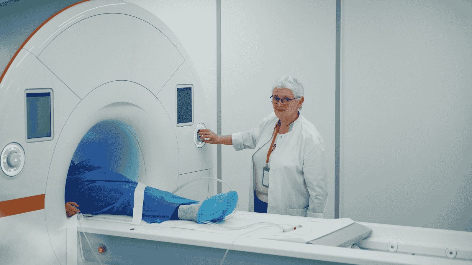
Dynamic Pelvis MRI and MR Defecography
The pelvic floor is a region at the lower part of the body, consisting of bones, muscles, and various soft tissues, where crucial body functions occur. This area houses organs such as the bladder, rectum, uterus, ovaries, and prostate gland. When structures in this area are damaged, it leads to changes in important functions like urinary retention and control of defecation. With an aging population, the quality of life can be negatively affected.
Obesity, previous surgical operations, constipation, trauma, pregnancy and childbirth, and intense physical exercise are among the factors that can cause damage to the muscles and other anatomical structures in this region, leading to chronic problems. These issues may include urinary problems like painful and/or frequent urination, constipation or fecal incontinence, prolapse of pelvic organs, pain and pressure sensations, spasms, and painful intercourse in women.
Clinical evaluation of discomfort and functional impairments in this region is quite challenging. Dynamic pelvis MRI and defecography play a significant role in diagnosing and treating disorders of this region.
Why is it needed?
- Cystocele
- Urethrocele
- Urethral hypermobility
- Uterovaginal prolapse
- Peritoneocele, enterocoele, and sigmoidocoele
- Rectocele
- Rectal intussusception and prolapse
- Pelvic floor weakness
- Dyssynergic defecation (prolonged absence of defecation)
- Anal incontinence
- Irritable bowel syndrome
Advanced MRI Applications
- Whole Body MRI
- Cardiac MRI
- Multiparametric Prostate MRI
- Magnetic Resonance Angiography (MRA)
- Prostate MRI
- Breast MRI
- Functional MRI (fMRI)
- Dynamic Pelvic MRI and MR Defecography
- Cranial (Brain, Head, Neck) MRI
- Abdominal and Pelvic MRI
- MRI Enterography